408-854-1883 starts at $30 per hr home care

Skip to main content

Affordable in home care | starts at $28 per hr

A New Model For How Brain Reward Response May Impact Anorexia

A New Model For How Brain Reward Response May Impact Anorexia

Summary: Researchers report the brain’s response to taste stimuli is linked to higher anxiety and a drive for thinness in those with anorexia.

Source: University of Colorado.

Researchers at the University of Colorado Anschutz Medical Campus have found that the brain’s response to taste stimuli is linked to high anxiety and a drive for thinness that could play a role in driving anorexia nervosa.

The study was published last week in the journal JAMA Psychiatry.

The researchers, led by Dr. Guido Frank, MD, associate professor of psychiatry and neuroscience at the University of Colorado School of Medicine, monitored a large group of patients with anorexia nervosa as they tasted sugar during brain imaging.

They found their brain response was higher than those in the control group, representing a biological marker for the illness. At the same time, this brain response was related to high anxiety and less weight gain for those being treated for anorexia nervosa.

Frank found that as these patients restricted their diet, a brain reward circuit associated with the neurotransmitter dopamine becomes more active but also triggers anxiety. This makes food avoidance worse and perpetuates the often deadly disease.

“When you lose weight your brain reward response goes up,” said Frank. “But instead of driving eating, we believe it elevates anxiety in anorexia nervosa, which makes them want to restrict more. This becomes then a vicious cycle.”

Using brain scans, the researchers examined 56 female adolescent and young adults with anorexia nervosa between the ages of 11 and 21 and 52 healthy control participants of the same age. They all learned to associate colored shapes with either getting or not getting a sugary solution. Sometimes when they expected sugar they got nothing, and sometimes when they didn’t expect sugar they received it.

Those with the eating disorder responded more strongly to the unexpected getting or not getting of sugar water, perhaps due to the release of dopamine.

The researchers found that the higher the brain response, the higher the harm avoidance in those with anorexia nervosa was. Harm avoidance is an anxiety measure for excessive worrying and fearfulness. In these patients, it pushes the drive for thinness and furthers body dissatisfaction.

Frank discovered that the higher the brain response, the lower the weight gain during treatment.

This brain reward response acted on the hypothalamus, which stimulates eating, in the anorexia nervosa group. The researchers hypothesized that this could make it possible to override and fend off signals to eat.

“An enhanced dopamine reward system response is an adaptation to starvation,” the study said. “Individuals vulnerable to developing anorexia nervosa could be particularly sensitive to food restriction and adaptations of reward response during the [mid-adolescence] development period.”

a scales

According to Frank, anorexia nervosa behavior could alter the brain circuits and impact its taste-reward processing mechanisms. Those who are already worried about shape and weight become even more concerned. And a strong response that says “feed me” might be overwhelming and trigger more food restriction instead of eating.

The study noted that while most people like sweet tasting things, those with eating disorders associate the taste with weight gain and try to avoid it. Frank found that the brain activation among the anorexia group was inversely connected with any pleasant experience of eating sugar.

“Our data raise the possibility that adolescents with anorexia nervosa in this study were negatively conditioned to sweet taste and may have developed an inverse association with dopamine release across the larger (brain) reward circuitry,” the study said.

Frank believes these insights could lead to new treatments for eating disorders.

“I hope we can use these findings to manipulate these biomarkers and design better treatments for this often-deadly disease,” he said.

ABOUT THIS NEUROSCIENCE RESEARCH ARTICLE

Source: David Kelly – University of Colorado
Publisher: Organized by NeuroscienceNews.com.
Image Source: NeuroscienceNews.com image is in the public domain.
Original Research: Open access research for “Association of Brain Reward Learning Response With Harm Avoidance, Weight Gain, and Hypothalamic Effective Connectivity in Adolescent Anorexia Nervosa” by Guido K. W. Frank, MD; Marisa C. DeGuzman, BA, BS; Megan E. Shott, BS; Mark L. Laudenslager, PhD; Brogan Rossi, BS; and Tamara Pryor, PhD in JAMA Psychiatry. Published July 19 2018.
doi:10.1001/jamapsychiatry.2018.2151

CITE THIS NEUROSCIENCENEWS.COM ARTICLE
University of Colorado”A New Model For How Brain Reward Response May Impact Anorexia.” NeuroscienceNews. NeuroscienceNews, 26 July 2018.
<http://neurosciencenews.com/reward-system-anorexia-9617/&gt;.

Abstract

Association of Brain Reward Learning Response With Harm Avoidance, Weight Gain, and Hypothalamic Effective Connectivity in Adolescent Anorexia Nervosa

Importance
Anorexia nervosa (AN) is associated with adolescent onset, severe low body weight, and high mortality as well as high harm avoidance. The brain reward system could have an important role in the perplexing drive for thinness and food avoidance in AN.

Objective
To test whether brain reward learning response to taste in adolescent AN is altered and associated with treatment response, striatal-hypothalamic connectivity, and elevated harm avoidance.

Design, Setting, and Participants
In this cross-sectional multimodal brain imaging study, adolescents and young adults with AN were matched with healthy controls at a university brain imaging facility and eating disorder treatment program. During a sucrose taste classical conditioning paradigm, violations of learned associations between conditioned visual and unconditioned taste stimuli evoked the dopamine-related prediction error (PE). Dynamic effective connectivity during sweet taste receipt was studied to investigate hierarchical brain activation across the brain network that regulates eating. The study was conducted from July 2012 to May 2017, and data were analyzed from June 2017 to December 2017.

Main Outcomes and Measures
Prediction error brain reward response across the insula, caudate, and orbitofrontal cortex; dynamic effective connectivity between hypothalamus and ventral striatum; and treatment weight gain, harm avoidance scores, and salivary cortisol levels and their correlations with PE brain response.

Results
Of 56 female participants with AN included in the study, the mean (SD) age was 16.6 (2.5) years, and the mean (SD) body mass index (BMI; calculated as weight in kilograms divided by height in meters squared) was 15.9 (0.9); of 52 matched female controls, the mean (SD) age was 16.0 (2.8) years, and the mean (SD) BMI was 20.9 (2.1). Prediction error response was elevated in participants with AN in the caudate head, nucleus accumbens, and insula (multivariate analysis of covariance: Wilks λ, 0.707; P = .02; partial η2 = 0.296), which correlated negatively with sucrose taste pleasantness. Bilateral AN orbitofrontal gyrus rectus PE response was positively correlated with harm avoidance (right ρ, 0.317; 95% CI, 0.091 to 0.539; P < .02; left ρ, 0.336; 95% CI, 0.112 to 0.550; P < .01) but negatively correlated with treatment BMI change (right ρ, −0.282; 95% CI, −0.534 to −0.014; P < .04; left ρ, −0.268; 95% CI, −0.509 to −0.018; P < .045). Participants with AN showed effective connectivity from ventral striatum to hypothalamus, and connectivity strength was positively correlated with insula and orbitofrontal PE response. Right frontal cortex PE response was associated with cortisol, which correlated with body dissatisfaction.

Conclusions and Relevance
These results further support elevated PE signal in AN and suggest a link between PE and elevated harm avoidance, brain connectivity, and weight gain in AN. Prediction error may have a central role in adolescent AN in driving anxiety and ventral striatal-hypothalamus circuit-controlled food avoidance.

Accumulation of toxic protein linked to mental health issues

System that Could Reduce Neurodegeneration in Huntington’s Discovered

Summary: Researchers have identified a mechanism that may reduce the toxic aggregation of the huntingtin protein. The findings could lead to new treatment options for Huntington’s patients.

Source: University of Cologne.

Dr David Vilchez and his team at CECAD have made an important step towards understanding the mechanisms that cause the neurodegenerative disorder Huntington’s disease. Particularly, they identified a system blocking the accumulation of toxin protein aggregates, which are responsible for neurodegeneration. The results have now been published in the journal ‘Nature Communications’.

Huntington’s disease is a neurodegenerative disorder that results in the death of brain cells, leading to uncontrolled body movement, loss of speech and psychosis. Mutations in the huntingtin gene cause the disease, resulting in the toxic aggregation of the huntingtin protein. The accumulation of these aggregates causes neurodegeneration and usually leads to the patient’s death within twenty years after the onset of the disease.

To examine the mechanisms underlying Huntington’s disease, Vilchez and his team used so-called induced pluripotent stem cells (iPSC) from Huntington’s disease patients, which are able to differentiate into any cell type, such as neurons. Induced pluripotent stem cells derived from patients with Huntington’s disease exhibit a striking ability to avoid the accumulation of toxic protein aggregates, a hallmark of the disease. Even though iPSCs express the mutant gene responsible for Huntington’s disease, no aggregates were found.

The protein degradation process

Ribbon diagram of ubiquitin, the highly conserved protein that serves as a molecular tag targeting proteins for degradation by the proteasome

Ubiquitination and targeting

Proteins are targeted for degradation by the proteasome with covalent modification of a lysine residue that requires the coordinated reactions of three enzymes. In the first step, a ubiquitin-activating enzyme (known as E1) hydrolyzes ATP and adenylylates a ubiquitin molecule. This is then transferred to E1’s active-site cysteine residue in concert with the adenylylation of a second ubiquitin.[41] This adenylylated ubiquitin is then transferred to a cysteine of a second enzyme, ubiquitin-conjugating enzyme (E2). In the last step, a member of a highly diverse class of enzymes known as ubiquitin ligases (E3) recognizes the specific protein to be ubiquitinated and catalyzes the transfer of ubiquitin from E2 to this target protein. A target protein must be labeled with at least four ubiquitin monomers (in the form of a polyubiquitin chain) before it is recognized by the proteasome lid.[42] It is therefore the E3 that confers substrate specificity to this system.[43] The number of E1, E2, and E3 proteins expressed depends on the organism and cell type, but there are many different E3 enzymes present in humans, indicating that there is a huge number of targets for the ubiquitin proteasome system.

The mechanism by which a polyubiquitinated protein is targeted to the proteasome is not fully understood. Ubiquitin-receptor proteins have an N-terminal ubiquitin-like (UBL) domain and one or more ubiquitin-associated (UBA) domains. The UBL domains are recognized by the 19S proteasome caps and the UBA domains bind ubiquitin via three-helix bundles. These receptor proteins may escort polyubiquitinated proteins to the proteasome, though the specifics of this interaction and its regulation are unclear.[44]

The ubiquitin protein itself is 76 amino acids long and was named due to its ubiquitous nature, as it has a highly conserved sequence and is found in all known eukaryotic organisms.[45] The genes encoding ubiquitin in eukaryotes are arranged in tandem repeats, possibly due to the heavy transcription demands on these genes to produce enough ubiquitin for the cell. It has been proposed that ubiquitin is the slowest-evolving protein identified to date.[46] Ubiquitin contains seven lysine residues to which another ubiquitin can be ligated, resulting in different types of polyubiquitin chains.[47] Chains in which each additional ubiquitin is linked to lysine 48 of the previous ubiquitin have a role in proteasome targeting, while other types of chains may be involved in other processes.[48][49]

The ubiquitination pathway

Unfolding and translocation

After a protein has been ubiquitinated, it is recognized by the 19S regulatory particle in an ATP-dependent binding step.[25] The substrate protein must then enter the interior of the 20S particle to come in contact with the proteolytic active sites. Because the 20S particle’s central channel is narrow and gated by the N-terminal tails of the α ring subunits, the substrates must be at least partially unfolded before they enter the core. The passage of the unfolded substrate into the core is called translocation and necessarily occurs after deubiquitination.[25] However, the order in which substrates are deubiquitinated and unfolded is not yet clear.[50] Which of these processes is the rate-limiting step in the overall proteolysis reaction depends on the specific substrate; for some proteins, the unfolding process is rate-limiting, while deubiquitination is the slowest step for other proteins.[24] The extent to which substrates must be unfolded before translocation is not known, but substantial tertiary structure, and in particular nonlocal interactions such as disulfide bonds, are sufficient to inhibit degradation.[51] The presence of intrinsically disordered protein segments of sufficient size, either at the protein terminus or internally, has also been proposed to facilitate efficient initiation of degradation.[52][53]

The gate formed by the α subunits prevents peptides longer than about four residues from entering the interior of the 20S particle. The ATP molecules bound before the initial recognition step are hydrolyzed before translocation. While energy is needed for substrate unfolding, it is not required for translocation.[24][25] The assembled 26S proteasome can degrade unfolded proteins in the presence of a non-hydrolyzable ATP analog, but cannot degrade folded proteins, indicating that energy from ATP hydrolysis is used for substrate unfolding.[24] Passage of the unfolded substrate through the opened gate occurs via facilitated diffusion if the 19S cap is in the ATP-bound state.[54]

The mechanism for unfolding of globular proteins is necessarily general, but somewhat dependent on the amino acid sequence. Long sequences of alternating glycine and alanine have been shown to inhibit substrate unfolding, decreasing the efficiency of proteasomal degradation; this results in the release of partially degraded byproducts, possibly due to the decoupling of the ATP hydrolysis and unfolding steps.[55] Such glycine-alanine repeats are also found in nature, for example in silk fibroin; in particular, certain Epstein–Barr virusgene products bearing this sequence can stall the proteasome, helping the virus propagate by preventing antigen presentation on the major histocompatibility complex.[56]
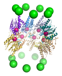
A cutaway view of the proteasome 20S core particle illustrating the locations of the active sites. The α subunits are represented as green spheres and the β subunits as protein backbones colored by individual polypeptide chain. The small pink spheres represent the location of the active-site threonine residue in each subunit. Light blue chemical structures are the inhibitor bortezomib bound to the active sites.

Proteolysis

The mechanism of proteolysis by the β subunits of the 20S core particle is through a threonine-dependent nucleophilic attack. This mechanism may depend on an associated water molecule for deprotonation of the reactive threonine hydroxyl. Degradation occurs within the central chamber formed by the association of the two β rings and normally does not release partially degraded products, instead reducing the substrate to short polypeptides typically 7–9 residues long, though they can range from 4 to 25 residues, depending on the organism and substrate. The biochemical mechanism that determines product length is not fully characterized.[57] Although the three catalytic β subunits have a common mechanism, they have slightly different substrate specificities, which are considered chymotrypsin-like, trypsin-like, and peptidyl-glutamyl peptide-hydrolyzing (PHGH)-like. These variations in specificity are the result of interatomic contacts with local residues near the active sites of each subunit. Each catalytic β subunit also possesses a conserved lysine residue required for proteolysis.[19]

Although the proteasome normally produces very short peptide fragments, in some cases these products are themselves biologically active and functional molecules. Certain transcription factors regulating the expression of specific genes, including one component of the mammalian complex NF-κB, are synthesized as inactive precursors whose ubiquitination and subsequent proteasomal degradation converts them to an active form. Such activity requires the proteasome to cleave the substrate protein internally, rather than processively degrading it from one terminus. It has been suggested that long loops on these proteins’ surfaces serve as the proteasomal substrates and enter the central cavity, while the majority of the protein remains outside.[58] Similar effects have been observed in yeast proteins; this mechanism of selective degradation is known as regulated ubiquitin/proteasome dependent processing (RUP).[59]

Ubiquitin-independent degradation

Although most proteasomal substrates must be ubiquitinated before being degraded, there are some exceptions to this general rule, especially when the proteasome plays a normal role in the post-translational processing of the protein. The proteasomal activation of NF-κB by processing p105 into p50 via internal proteolysis is one major example.[58] Some proteins that are hypothesized to be unstable due to intrinsically unstructured regions,[60] are degraded in a ubiquitin-independent manner. The most well-known example of a ubiquitin-independent proteasome substrate is the enzyme ornithine decarboxylase.[61] Ubiquitin-independent mechanisms targeting key cell cycle regulators such as p53 have also been reported, although p53 is also subject to ubiquitin-dependent degradation.[62] Finally, structurally abnormal, misfolded, or highly oxidized proteins are also subject to ubiquitin-independent and 19S-independent degradation under conditions of cellular stress.[63]

Evolution

The assembled complex of hslV(blue) and hslU (red) from E. coli. This complex of heat shock proteins is thought to resemble the ancestor of the modern proteasome.

The 20S proteasome is both ubiquitous and essential in eukaryotes. Some prokaryotes, including many archaea and the bacterial order Actinomycetales also share homologs of the 20S proteasome, whereas most bacteria possess heat shock genes hslV and hslU, whose gene products are a multimeric protease arranged in a two-layered ring and an ATPase.[64] The hslV protein has been hypothesized to resemble the likely ancestor of the 20S proteasome.[65] In general, HslV is not essential in bacteria, and not all bacteria possess it, whereas some protists possess both the 20S and the hslV systems.[64]Many bacteria also possess other homologs of the proteasome and an associated ATPase, most notably ClpP and ClpX. This redundancy explains why the HslUV system is not essential.

Sequence analysis suggests that the catalytic β subunits diverged earlier in evolution than the predominantly structural α subunits. In bacteria that express a 20S proteasome, the β subunits have high sequence identity to archaeal and eukaryotic β subunits, whereas the α sequence identity is much lower. The presence of 20S proteasomes in bacteria may result from lateral gene transfer, while the diversification of subunits among eukaryotes is ascribed to multiple gene duplication events.[64]

Cell cycle control

Cell cycle progression is controlled by ordered action of cyclin-dependent kinases (CDKs), activated by specific cyclins that demarcate phases of the cell cycle. Mitotic cyclins, which persist in the cell for only a few minutes, have one of the shortest life spans of all intracellular proteins.[1] After a CDK-cyclin complex has performed its function, the associated cyclin is polyubiquitinated and destroyed by the proteasome, which provides directionality for the cell cycle. In particular, exit from mitosis requires the proteasome-dependent dissociation of the regulatory component cyclin B from the mitosis promoting factor complex.[66] In vertebrate cells, “slippage” through the mitotic checkpoint leading to premature M phase exit can occur despite the delay of this exit by the spindle checkpoint.[67]

Earlier cell cycle checkpoints such as post-restriction point check between G1 phase and S phase similarly involve proteasomal degradation of cyclin A, whose ubiquitination is promoted by the anaphase promoting complex (APC), an E3 ubiquitin ligase.[68] The APC and the Skp1/Cul1/F-box protein complex (SCF complex) are the two key regulators of cyclin degradation and checkpoint control; the SCF itself is regulated by the APC via ubiquitination of the adaptor protein, Skp2, which prevents SCF activity before the G1-S transition.[69]

Individual components of the 19S particle have their own regulatory roles. Gankyrin, a recently identified oncoprotein, is one of the 19S subcomponents that also tightly binds the cyclin-dependent kinase CDK4 and plays a key role in recognizing ubiquitinated p53, via its affinity for the ubiquitin ligase MDM2. Gankyrin is anti-apoptotic and has been shown to be overexpressed in some tumor cell types such as hepatocellular carcinoma.[70]

Regulation of plant growth

In plants, signaling by auxins, or phytohormones that order the direction and tropism of plant growth, induces the targeting of a class of transcription factor repressors known as Aux/IAA proteins for proteasomal degradation. These proteins are ubiquitinated by SCFTIR1, or SCF in complex with the auxin receptor TIR1. Degradation of Aux/IAA proteins derepresses transcription factors in the auxin-response factor (ARF) family and induces ARF-directed gene expression.[71] The cellular consequences of ARF activation depend on the plant type and developmental stage, but are involved in directing growth in roots and leaf veins. The specific response to ARF derepression is thought to be mediated by specificity in the pairing of individual ARF and Aux/IAA proteins.[72]

Apoptosis

Both internal and external signals can lead to the induction of apoptosis, or programmed cell death. The resulting deconstruction of cellular components is primarily carried out by specialized proteases known as caspases, but the proteasome also plays important and diverse roles in the apoptotic process. The involvement of the proteasome in this process is indicated by both the increase in protein ubiquitination, and of E1, E2, and E3 enzymes that is observed well in advance of apoptosis.[73][74][75] During apoptosis, proteasomes localized to the nucleus have also been observed to translocate to outer membrane blebs characteristic of apoptosis.[76]

Proteasome inhibition has different effects on apoptosis induction in different cell types. In general, the proteasome is not required for apoptosis, although inhibiting it is pro-apoptotic in most cell types that have been studied. Apoptosis is mediated through disrupting the regulated degradation of pro-growth cell cycle proteins.[77] However, some cell lines — in particular, primary cultures of quiescent and differentiated cells such as thymocytes and neurons — are prevented from undergoing apoptosis on exposure to proteasome inhibitors. The mechanism for this effect is not clear, but is hypothesized to be specific to cells in quiescent states, or to result from the differential activity of the pro-apoptotic kinase JNK.[78] The ability of proteasome inhibitors to induce apoptosis in rapidly dividing cells has been exploited in several recently developed chemotherapy agents such as bortezomib and salinosporamide A.

Response to cellular stress

In response to cellular stresses – such as infectionheat shock, or oxidative damage – heat shock proteins that identify misfolded or unfolded proteins and target them for proteasomal degradation are expressed. Both Hsp27 and Hsp90chaperone proteins have been implicated in increasing the activity of the ubiquitin-proteasome system, though they are not direct participants in the process.[79] Hsp70, on the other hand, binds exposed hydrophobic patches on the surface of misfolded proteins and recruits E3 ubiquitin ligases such as CHIP to tag the proteins for proteasomal degradation.[80] The CHIP protein (carboxyl terminus of Hsp70-interacting protein) is itself regulated via inhibition of interactions between the E3 enzyme CHIP and its E2 binding partner.[81]

Similar mechanisms exist to promote the degradation of oxidatively damaged proteins via the proteasome system. In particular, proteasomes localized to the nucleus are regulated by PARP and actively degrade inappropriately oxidized histones.[82] Oxidized proteins, which often form large amorphous aggregates in the cell, can be degraded directly by the 20S core particle without the 19S regulatory cap and do not require ATP hydrolysis or tagging with ubiquitin.[63] However, high levels of oxidative damage increases the degree of cross-linking between protein fragments, rendering the aggregates resistant to proteolysis. Larger numbers and sizes of such highly oxidized aggregates are associated with aging.[83]

Dysregulation of the ubiquitin proteasome system may contribute to several neural diseases. It may lead to brain tumors such as astrocytomas.[84] In some of the late-onset neurodegenerative diseases that share aggregation of misfolded proteins as a common feature, such as Parkinson’s disease and Alzheimer’s disease, large insoluble aggregates of misfolded proteins can form and then result in neurotoxicity, through mechanisms that are not yet well understood. Decreased proteasome activity has been suggested as a cause of aggregation and Lewy body formation in Parkinson’s.[85] This hypothesis is supported by the observation that yeast models of Parkinson’s are more susceptible to toxicity from α-synuclein, the major protein component of Lewy bodies, under conditions of low proteasome activity.[86] Impaired proteasomal activity may underlie cognitive disorders such as the autism spectrum disorders, and muscle and nerve diseases such as inclusion body myopathy.[84]

Role in the immune system

The proteasome plays a straightforward but critical role in the function of the adaptive immune system. Peptide antigens are displayed by the major histocompatibility complex class I (MHC) proteins on the surface of antigen-presenting cells. These peptides are products of proteasomal degradation of proteins originated by the invading pathogen. Although constitutively expressed proteasomes can participate in this process, a specialized complex composed of proteins, whose expression is induced by interferon gamma, are the primary producers of peptides which are optimal in size and composition for MHC binding. These proteins whose expression increases during the immune response include the 11S regulatory particle, whose main known biological role is regulating the production of MHC ligands, and specialized β subunits called β1i, β2i, and β5i with altered substrate specificity. The complex formed with the specialized β subunits is known as the immunoproteasome.[15] Another β5i variant subunit, β5t, is expressed in the thymus, leading to a thymus-specific “thymoproteasome” whose function is as yet unclear.[87]

The strength of MHC class I ligand binding is dependent on the composition of the ligand C-terminus, as peptides bind by hydrogen bonding and by close contacts with a region called the “B pocket” on the MHC surface. Many MHC class I alleles prefer hydrophobic C-terminal residues, and the immunoproteasome complex is more likely to generate hydrophobic C-termini.[88]

Due to its role in generating the activated form of NF-κB, an anti-apoptotic and pro-inflammatory regulator of cytokine expression, proteasomal activity has been linked to inflammatory and autoimmune diseases. Increased levels of proteasome activity correlate with disease activity and have been implicated in autoimmune diseases including systemic lupus erythematosus and rheumatoid arthritis.[15]

The proteasome is also involved in Intracellular antibody-mediated proteolysis of antibody-bound virions. In this neutralisation pathway, TRIM21 (a protein of the tripartite motif family) binds with immunoglobulin G to direct the virion to the proteasome where it is degraded.


Tip: Kill the virus in your brain with whole foods , adequate sleep and exercise in the sun.


Soda and diabetes risk

Soda kills mice

Telemedicine and the Coming Physician Shortage

Telemedicine and the Coming Physician Shortage
With an aging population and a limited supply of physicians, the shortage is likely unavoidable. But by increasing the use of telemedicine and non-physician clinicians, and improving care coordination and chronic care management, we can reduce reliance on traditional doctor visits. Download Here
Teladoc Eyes Global Telehealth Network With Advance Medical Deal
Teladoc’s purchase of Spain-based Advance Medical gives the telehealth company an inroads to the lucrative Asian and Latin American markets and continues a string of high-profile partnerships in the digital health industry.
Full Story
Headlines
Using Telehealth, mHealth Technology to Help Seniors Age in Place
Picking The Right Telehealth Platform For a Small or Solo Practice
Telehealth, mHealth Make Nurses Pivotal Presence in Healthcare
Featured Resource
How to Reduce Preventable Readmissions with Healthcare IT
As 2,610 healthcare providers face readmission penalties this year, the quest for efficient readmission-reducing strategies becomes paramount. How can branded hospital apps help reduce preventable readmissions and curb the healthcare costs?
Download Here
LAST CHANCE
Telehealth2018
June 7-8, 2018 | San Diego, CA
A Must-Attend You Won’t Want to Miss!

Creating a Culture of Support for Telehealth: Gain first-hand experiences and real-world advice from leaders atPartners, Methodist Health System,and Mount Sinai, about how to achieve buy-in from all levels of your organization by developing clear clinical and business objectives, and a solid strategy for meeting them

FINAL CHANCE! Registration Closes TODAY JUNE 6TH!
As a valued member of mHealthIntelligence REGISTER FOR $399! – Use Code: MHEALTH
*For Providers & Payers Only – Not Valid on Previous Registrations*

Register Here
Featured Resource
How Millennials are Redefining Healthcare
Millennials are quickly becoming the dominant healthcare consumers, both for themselves, their families and as caregivers. Having grown up in a technology-first environment, however, they are increasingly dissatisfied with the static nature of healthcare options and are seeking out alternatives.
Download Here
St. Joseph’s Villa Uses Telehealth to Connect With Children in Crisis
Virginia-based St. Joseph’s Villa has added a telehealth service to its 8-bed crisis stabilization unit, giving adolescents an instant connection to specialists. Officials say the virtual visits have improved outcomes. Full Story
mHealth Finds a Place for Fitbit in Remote Patient Monitoring Research
Cedars-Sinai and UCLA researchers have found that a consumer-grade mHealth wearable – in this case, a Fitbit – can help providers in remote patient monitoring programs for patients with heart issues. Full Story
Jefferson Health Tests an mHealth Wearable for Pneumonia Detection
Researchers at Jefferson Health’s Sidney Kimmel Cancer Center are studying whether an mHealth wearable used to monitor breathing in asthma patients can be adapted to detect signs of pneumonia in patients with lung cancer. Full Story
Featured Resources
10 Best Practices for Discovering the Best Mobile and IoT Devices for Healthcare
5 Common Missteps on the Path to Enterprise Mobility
Where Compassion and Technology Meet: Children’s Mercy
Everything Urgent Cares Need to Know to Improve Patient Engagement
Texting Your Patients: Why It’s Hard and How to Get it Right
How to Stop Making Patients Pay the Price for Poor Communications

Top health hacks 7-26-2018

View
Eggplant and apple cider vinegar for skin cancer
View
Non pasteurized beers have more health benefits
View
Home page / Archives
View
Apple cider vinegar kills parasites, cleansing to the liver and prevents stroke
View
DMSO, hydrogen peroxide and Vit C fight cancer cells
View
Signs of the preactive/ active phase of dying and medications for terminally ill
View
Philippines Coconut Wine -Tuba
View
Can Gout be cured permanently?
View
Nitric Oxide Dump Exercise with nose breathing to lower blood pressure and thin blood
View
Foods to eat and avoid when you have Gout and leg pains
View
Can balsamic vinegar help with gout?
View
Massage oil of fresh ginger and coconut oil relieves joint pain
View
MEDICATIONS TO AVOID that worse PD (Parkinson’s disease)
View
MSM powder benefits – Alzheimer is a sulfur deficiency
View
Fatigue and Red (bloodshot) eyes from WebMD
View
Anti-aging and Parkinson/Alzheimer’s prevention: Enzymes and apple cider vinegar
View
What’s a good analogy to explain the immune system?
View
Fruits and leaves of Figs as anti-cancer
View
Hiatal Hernia, Pancreatitis, Pancreatic Cancer and the Western Diet
View
Increase the body’s oxygen carrying capacity with exercise, EPO and whole foods
View
What the Latest Mueller Indictment Reveals About WikiLeaks’ Ties to Russia—and What It Doesn’t
View
Avoid chronic bronchitis with green apple, onions, garlic, vinegar and rest
View
About fingerprinting, IHSS and worker’s comp in California for home care
View
Toxicology test for pregnant women
View
2
Viral infection affects the vascular system and the brain
View
2
Philippines president Dutarte asked each town to prepare a list of drug users and pushers
View
2
How the digestive tract communicates with the brain
View
2
Eat protein-rich food when drinking alcohol to protect your stomach
View
2
Disease prediction with HELO wearable, own a piece of the market
View
2
Chlorine rich foods are cleansing (avocado, coconut, onions, parsnips)
View
1
Antisocial personality disorder in 70% of prison inmates
View
1
Apple cider vinegar for knee swelling and more uses by Dr Mercola
View
1
Mullein herb for lung and breast health – COPD signs, symptoms and diagnosis
View
1
Detox your lungs from air pollution and metal toxins and for early lung cancer
View
1
Liver cleanse to help your vision and memory
View
1
Digestive enzymes help in healing fractures, preventing kidney stones and heart disease and more
View
1
Leaky gut, leaky brain, eat your garlic and pickles by C Guthrie
View
1
Restore your vision naturally y Dr. Mercola
View
1
IRS-1 protein in blood, indicative of Alzheimer
View
1
Boron fights radiation by Dr Mercola
View
1
Anaemia, causes, drugs, folate in beer, beer carbs
View
1
Fasting, sun bathing ,Vit C, Lysine, turmeric, green tea, carrots and raw food diet to reduce tumor size
View
1
Burn Fat by knocking out the gene that produces folliculin in fat cells, thru exercise and eating Vitamin C and iron-rich foods
View
1
Gov. Brown signs bill that limits seizure of assets of many Medi-Cal recipients
View
1
How long does dexedrine stay in your system?
View
1
Can percumin prevent Parkinson’s disease?
View
1
Dry mouth and bad breath by Dr Mercola
View
1
Can blunt force trauma cause brain cancer?
View
1
Lung Cancer deaths from 55-70
View
1
10 blood tests for your physical check up
View
1
Hemochromatosis patients should be on a low-iron diet
View
1
Vagus nerve stimulation thru breathing, laughs and yoga
View
1
Parasites and their effects on your immune system
View
1
Lung cancer in the Philippines
View
1
Gastroparesis, Betain HCL, diabetes and stomach health
View
1
Foods that delay the rate of brain atrophy in patients with Alzheimer’s disease
View
1
Loss of smell and neurodegenerative disease
View
1
Top posts in November
View
1
In Sympathetic Nervous System Why the digestion of food is slow? Where as in fight or flight we need more energy.?
View
1
Lung cancer and heavy metal toxins
View
1
Iron and sweet wormwood herb kill breast cancer cells in 16hrs
View
1
Reducing Failures or Defects in Medical Device a Corrective and Preventive Action Plan
View
1
Why is pizza bad for cholesterol?
View
1
Surviving prostate cancer by Dr Mercola
View
1
Argentinge Tango in San Jose CA
View
1
The Von Trump Family Singers
View
1
Slow down, keep a healthy weight and deep breathing to prevent stroke
View
1
Symptoms of Crohn’s disease, LRRK2 genes , Parkinson and nutrition
View
1
20 renowned US-China investors
View
1
Baking soda for mosquito bites
View
1
Top viewed posts for June 2017
View
1
Herbal oil for Lice, Scabies and skin issues
View
1
French startup launches hydrogen-powered bicycles
View
1
Misdiagnosed thyroid cancers by Dr Mercola
View
1
Alzheimer’s protein may spread like an infection, human brain scans suggest
View
1
Bone spurs, free calcium and Dementia
View
1
Chest pain, flu and fever
View
1
FDA and telehealth
View
1
Prostate flush and masturbation
View
1
Washington Post: Opinion section
View
1
Volunteer as caregiver for health benefits and tuition assistance
View
1
Benefits of activated charcoal by Dr Axe
View
1
Binge eating and Rhodiola rosea
View
1
Fungus , raw carrots and prostate cancer
View
1
A Theory of Trump Kompromat
View
1
Inflammation to colitis to Alzheimer’s disease
View
1
Coconut and cinnamon for Alzheimer’s disease
View
1
New Study Discovers ‘Killer Peptide’ That Helps Eliminate Resistant Cancer Cells
View
1
Nitric Oxide for strong blood vessels’ cells , up with exercise, melons, cucumber, Vit C, E, amino acid – L-arginine, L-citrulline
View
1
News Medical Life Sciences
View
1
Prostate health, intensive nutrition and AGELOC Youth as anti-inflammatory
View
1
My passion in health education as seen at quora.com
View
1
ALOE FEROX plant extract (a laxative agent in South Africa) increased intestinal secretion and motility in constipated rats
View
1
Corporate ties to Customs and Border Protection (CBP)
View
1
SumOfUs at the “Together Against Trump” protest in London
View
1
Jim Jefferies on US healthcare
View
1
Single neurons regulate their overall action potential firing rate
View
1
Samsung and WellDoc partner to offer direct-to-consumer version of diabetes management app
View
1
Free classes in San Jose from Qigong to Yoga for bay area cancer patients at cancercarepoint.org
View
1
Interaction between men’s testes and the immune system
View
1
Cancer signs by Dr Mercola
View
1
What Causes Muscle Cramps and Spasms?
View
1
Washington Post 7-17-2017
View
1
Eurycoma longifolia – Tongkat Ali for strength and libido
View
1
Our ability to distinguish between what is real and what is perceived
View
1
NAC, activated charcoal , sleep and parasites
View
1
The Scent of Coffee Appears to Boost Math Performance
View
1
Baking soda , lemon and apple cider vinegar to repair kidney damage from sugar
View
1
Soda and Diabetes
View
1
Massive brain reorganization was reversed in a child with hand transplant
View
1
Cortisol can reduce cravings for those addicted to heroin
View
1
MRI SHOWS BRAIN DISRUPTIONS IN CHILDREN WITH PTSD
View
1
Grand multiparity was associated with high risk of Alzheimer’s
View
1
Autism Risk Determined by Health of Mother’s Gut
View
1
Neurons Can Carry More Than One Signal at a Time
View
1
Depression and Antidepressants Linked to Increased Risk of Venous Thrombo Embolism

Cognitive and Motor Training Combined May Slow or Reverse Dementia

Cognitive and Motor Training Combined May Slow or Reverse Dementia

Summary: A new study reports 30 minutes of cognitive and motor training once a week can slow the progression of, and possibly reverse, the symptoms of dementia.

Source: York University.

Researchers at York University’s Faculty of Health found that just 30 minutes of visually-guided movements per week can slow and even reverse the progress of dementia. Those in the early stages of dementia who were exposed to 30 minutes a week to a game which used rules to make visually-guided movements, were able to slow down the progress of dementia and for some, even reverse their cognitive function to healthy status.

Previous approaches have used cognitive training alone or aerobic exercise training alone. This study published in Dementia and Geriatric Disorders, is the first to investigate the impact of combining both types of approaches on cognitive function in elderly people with various degrees of cognitive defects.

“We found cognitive-motor integration training slows down the progress of dementia, and for those just showing symptoms of dementia, this training can actually revert them back to healthy status, stabilizing them functionally,” says lead researcher, Lauren Sergio, professor in the School of Kinesiology and Health Science and Centre for Vision Research at York University.

In the intervention study, a total of 37 elderly people located at senior centres, were divided into four groups based on their level of cognition. They completed a 16-week cognitive-motor training program that consisted of training sessions involving playing a videogame that required goal-directed hand movements on a computer tablet for 30 minutes a week. Before and after the training program, all participants completed a series of tests to establish their level of cognition and visuomotor skills. Sergio’s team performed tests to evaluate cognitive function 14 days prior to and after the intervention period, respectively. Her team observed an overall change in all groups and, specifically, a significant improvement in measures of overall cognition in the sub average cognition group and the mild-to-moderate cognitive deficits group. “These results suggest that even in the earliest stages of neurodegeneration, the aging brain has enough neuroplasticity left that if you can train it on this kind of thinking and moving task, it will improve their cognitive skills,” says Sergio. “The brain still possesses the functional capacities to form sufficient new synaptic connections to induce relevant changes on a systems level.”

Sergio adds the findings suggest that repetitive cognitive-motor integration training may in fact strengthen the involved neural networks and improve cognitive and functional abilities. Researchers believe the frontal lobe is ‘talking’ to the motor control areas and this is what is paving the way for success.

an old lady

The study further found that those in the severe cognitive deficits group who did 30 minutes of this eye-hand task did not decline in their cognitive deficits as expected, but instead stayed the same.

“Generally, you expect someone with severe dementia to have their cognitive function decline over five months, but in our study, they all stabilized.”

Sergio says the findings show promise for those who have early-stage dementia because the approach is easy to administer remotely and shows more promise than the basic cognitive training.

York University champions new ways of thinking that drive teaching and research excellence. Our students receive the education they need to create big ideas that make an impact on the world. Meaningful and sometimes unexpected careers result from cross-disciplinary programming, innovative course design and diverse experiential learning opportunities. York students and graduates push limits, achieve goals and find solutions to the world’s most pressing social challenges, empowered by a strong community that opens minds. York U is an internationally recognized research university – our 11 faculties and 25 research centres have partnerships with 200+ leading universities worldwide. Located in Toronto, York is the third largest university in Canada, with a strong community of 53,000 students, 7,000 faculty and administrative staff, and more than 300,000 alumni.

ABOUT THIS NEUROSCIENCE RESEARCH ARTICLE

Source: Anjum Nayyar – York University
Publisher: Organized by NeuroscienceNews.com.
Image Source: NeuroscienceNews.com image is in the public domain.
Original Research: Open access research for “Thinking-While-Moving Exercises May Improve Cognition in Elderly with Mild Cognitive Deficits: A Proof-of-Principle Study” by de Boer C., Echlin H.V., Rogojin A., Baltaretu B.R., Sergio L.E. in Dementia and Geriatric Disorders. Published July 10 2018.
doi:10.1159/000490173

CITE THIS NEUROSCIENCENEWS.COM ARTICLE
York University”Cognitive and Motor Training Combined May Slow or Reverse Dementia.” NeuroscienceNews. NeuroscienceNews, 25 July 2018.
<http://neurosciencenews.com/cognitive-motor-training-dementia-9609/&gt;.

Abstract

Thinking-While-Moving Exercises May Improve Cognition in Elderly with Mild Cognitive Deficits: A Proof-of-Principle Study

Background: Noninvasive interventions to aid healthy cognitive aging are considered an important healthcare priority. Traditional approaches typically focus on cognitive training or aerobic exercise training. In the current study, we investigate the effect of exercises that directly combine cognitive and motor functions on visuomotor skills and general cognition in elderly with various degrees of cognitive deficits.

Subjects and Methods: A total of 37 elderly, divided into four groups based on their level of cognition, completed a 16-week cognitive-motor training program. The weekly training sessions consisted of playing a videogame requiring goal-directed hand movements on a computer tablet for 30 minutes. Before and after the training program, all participants completed a test battery to establish their level of cognition and visuomotor skills.

Results: We observed an overall change in visuomotor behavior in all groups, as participants completed the tasks faster but less accurately. More importantly, we observed a significant improvement in measures of overall cognition in the subaverage cognition group and the mild-to-moderate cognitive deficits group.

Conclusion: Our findings indicate that (1) cognitive-motor exercises induce improved test scores, which is most prominent in elderly with only mild cognitive deficits, and (2) cognitive-motor exercises induce altered visuomotor behavior and slight improvements in measures of general cognition.

Widespread Connections Among Neurons Help the Brain Distinguish Smells

Widespread Connections Among Neurons Help the Brain Distinguish Smells

Summary: Researchers say the randomness of the piriform cortex plays a critical role when it comes to distinguishing between similar odors.

Source: Salk Institute.

Can you tell the smell of a rose from the scent of a lilac? If so, you have your brain’s piriform cortex to thank. Compared to many parts of the brain, the piriform cortex–which lets animals and humans process information about smells–looks like a messy jumble of connections between cells called neurons. Now, Salk Institute researchers have illuminated how the randomness of the piriform cortex is actually critical to how the brain distinguishes between similar odors.

“The standard paradigm is that information in the brain is encoded by which cells are active, but that’s not true for the olfactory system,” says Charles Stevens, Distinguished Professor Emeritus in Salk’s Molecular Neurobiology Laboratory and coauthor of the new work. “In the olfactory system, it turns out it’s not a matter of which cells are active, but how many cells are active and how active they are.”

Aside from better understanding how smells are processed, the new research, published in theJournal of Comparative Neurology on July 17, 2018, could also lead to greater insight into how some parts of the brain organize information.

When odorant molecules–the signature of any given smell–bind to the receptors in a person’s nose, the signal is transmitted to the olfactory bulb, and from there to the piriform cortex. In other sensory systems–like the visual system–information maintains a strict order as it moves through the brain. Particular parts of the eye, for instance, always transmit information to specific parts of the visual cortex. But researchers have long known that this order is missing in the piriform cortex.

“We haven’t been able to discern any order in the piriform cortex connections in any species,” says coauthor Shyam Srinivasan, an assistant project scientist at the University of California San Diego’s Kavli Institute for Brain and Mind. “Any given odor lights up about 10 percent of neurons that seem to be scattered all over the piriform cortex.”

To start working out the details of how the piriform cortex encodes odor information–and whether its connections are truly random–Stevens and Srinivasan analyzed the piriform cortices of nine mice using a variety of staining and microscopy techniques that let them visualize different cell types in the brain region. Their first goal: to quantify the number and density of cells in the piriform cortex.

“This was really like a survey,” explains Srinivasan. “We counted the cells in different representative areas and averaged them across the whole region.”

The mouse piriform cortex, they concluded, has around half a million neurons in it, divided equally between the larger, less dense posterior piriform and the smaller, more dense anterior piriform.

Using this initial information on density and neuron number, as well as knowledge from previous studies on the number of neurons in the olfactory bulb and how many neuronal connections–or synapses–connect the olfactory bulb to the piriform cortex, the pair of researchers was able to draw a surprising finding: each neuron in the olfactory bulb is connected to nearly every single neuron in the piriform cortex.

piriform cortex

“Every cell in the piriform is getting information from essentially every odor receptor there is,” says Stevens. “There’s not one ‘coffee smell’ neuron but a whole bunch of coffee cell neurons all over the place.” Rather than a single receptor detecting one odor and lighting up one cluster of telltale neurons, he explains, each odor has a fingerprint that’s based more on the strength of the connections–while the smell of coffee may activate nearly the same neurons in the piriform cortex as the smell of chocolate, they’ll activate each neuron to a different degree.

“One advantage to this system is that it can encode very complex information,” says Srinivasan. “It also makes it very robust to noise.” If one neuron sends a “noisy” signal–stronger or weaker activation than it should–the noise gets cancelled out by the many other neurons sending simultaneous, more accurate signals.

The researchers would like to repeat the work in other animals to see where similarities and differences lie. They also are interested in looking into other areas of the brain that have long been assumed to be dominated by seemingly random connections to see if they’re organized in the same way.

ABOUT THIS NEUROSCIENCE RESEARCH ARTICLE

Funding: The study was funded by the Kavli Institute for Brain and Mind at UC San Diego and the National Science Foundation.

Source: Salk Institute
Publisher: Organized by NeuroscienceNews.com.
Image Source: NeuroscienceNews.com image is credited to Salk Institute.
Original Research: Abstract for “The distributed circuit within the piriform cortex makes odor discrimination robust” by Shyam Srinivasan and Charles F Stevens in Journal of Comparative Neurology. Published July 17 2018.
doi:10.1002/cne.24492

CITE THIS NEUROSCIENCENEWS.COM ARTICLE
Salk Institute”Widespread Connections Among Neurons Help the Brain Distinguish Smells.” NeuroscienceNews. NeuroscienceNews, 25 July 2018.
<http://neurosciencenews.com/olfaction-networks-9610/&gt;.

Abstract

The distributed circuit within the piriform cortex makes odor discrimination robust

Distributed circuits wherein connections between subcircuit components seem to be randomly distributed are common to the olfactory circuit, hippocampus, and cerebellum. In such circuits, activation patterns seem random too, showing no detectable spatial preference, and contrast with regions that have topographic connections between sub‐circuits and topographic activation patterns. Quantitative studies of topographic circuits in the neocortex have yielded common principles of organization. Whether distributed circuits share similar principles of organization is unknown because similar quantitative information is missing and the way they encode information remains a challenge. We addressed these needs by providing a quantitative description of the mouse piriform cortex, a paleocortical distributed circuit that subserves olfaction. The quantitative information provided two insights. First, with a nearly parameter‐free model of the olfactory circuit, we show that the piriform cortex robustly maintains odor information and discrimination ability present in the olfactory bulb. Second, the paleocortex is quantitatively different from the neocortex: it has a lower surface area density, which decreases from the anterior to posterior paleocortex contrasting with the uniform neuronal density of the neocortex. These insights might also apply to other distributed circuits like the hippocampus.

Depression and Antidepressants Linked to Increased Risk of Venous Thrombo Embolism

Depression and Antidepressants Linked to Increased Risk of VTE

Summary: Researchers have identified a link between depression, antidepressant use and an increased risk of developing venous thromboembolism.

Source: University of Bristol.

In the first review of its kind, new research has found that depression and the use of antidepressants are each associated with an increased risk of venous thromboembolism (VTE). The study led by academics from the Musculoskeletal Research Unit at the University of Bristol has also shown that each of the various classes of antidepressant medications are associated with an increased risk of VTE.

VTE, a condition in which blood clots form in the veins of the legs or lungs, is a life-threatening condition and its treatment is associated with high healthcare costs. The research, published in Annals of Medicine, conducted a systematic review and meta-analysis of published observational studies evaluating the associations of depression and antidepressant use with VTE risk.

There have been reports that both depression and use of antidepressant drugs might be associated with an increased risk of VTE. These reports have, however, been conflicting. Previous studies have reported mixed results, some reporting evidence of associations and others reporting no evidence of associations. The researchers have clarified the evidence by bringing all published studies together.

Though the study could not prove if the observed findings are mainly driven by the antidepressant drugs or depression itself or both, it does show that a relationship exists between depression, antidepressant use, and VTE.

woman

Antidepressant medications have multiple indications, which include anxiety, pain, and neuralgia and their use is on the increase on a global scale. Given that VTE is a public health burden, the study’s findings highlight the need for prescribers and healthcare professionals to evaluate patients to determine their excess risk of VTE during their management.

Dr Setor Kunutsor, Research Fellow from the Musculoskeletal Research Unit at the Bristol Medical School: Translational Health Sciences (THS) and lead researcher, said: “These findings are very useful to me as both a clinician and a researcher. It gives me the information I need, especially when prescribing antidepressant medications to my patients.”

The research findings do not prove cause and effect and further studies are needed to show if the associations the study has demonstrated are causal and whether it is depression or antidepressant use or both which drives an increase in VTE risk. These would need to involve studies that are able to isolate depression from antidepressant medications. For example, researchers could assess if individuals who are not depressed but use antidepressants for a condition such as neurologic or gastrointestinal disease, are at an increased risk of VTE.

ABOUT THIS NEUROSCIENCE RESEARCH ARTICLE

Source: Joanne Fryer – University of Bristol
Publisher: Organized by NeuroscienceNews.com.
Image Source: NeuroscienceNews.com image is in the public domain.
Original Research: Abstract for “Depression, antidepressant use, and risk of venous thromboembolism: systematic review and meta-analysis of published observational evidence” by Setor K. Kunutsor, Samuel Seidu & Kamlesh Khunti in Annals of Medicine. Published July 12 2018.
doi:10.1080/07853890.2018.1500703

CITE THIS NEUROSCIENCENEWS.COM ARTICLE
University of Bristol”Depression and Antidepressants Linked to Increased Risk of VTE.” NeuroscienceNews. NeuroscienceNews, 25 July 2018.
<http://neurosciencenews.com/vte-depression-9611/&gt;.

Abstract

Depression, antidepressant use, and risk of venous thromboembolism: systematic review and meta-analysis of published observational evidence

Purpose: Evidence on the association between depression, antidepressant use and venous thromboembolism (VTE) risk is conflicting. We conducted a systematic review and meta-analysis of published observational studies evaluating the associations of depression and antidepressant use with VTE risk.

Design: Eligible studies were identified in a literature search of MEDLINE, Embase, Web of Science and reference list of relevant studies up to April 2018. Pooled relative risks (RRs) with 95% confidence intervals (CIs) were calculated aggregated using random effects models.

Results: Eight observational studies with data on 960 113 non-overlapping participants and 9027 VTE cases were included. The pooled RR (95% CI) for VTE comparing antidepressant use with no antidepressant use was 1.27 (1.06-1.51). Tricyclic antidepressants, selective serotonin reuptake inhibitors and other antidepressants were each associated with an increased VTE risk; 1.16 (1.06-1.27), 1.12 (1.02-1.23), and 1.59 (1.21-2.09) respectively. In pooled analysis of three studies that compared patients with depression versus individuals without depression, the RR for VTE was 1.31 (1.13-1.53).

Conclusion: Pooled observational evidence suggests that depression and use of antidepressants are each associated with an increased VTE risk. The effect of antidepressant drugs on VTE may be a class effect. The mechanistic pathways underlying these associations deserve further evaluation.

Kushner dodges subpoena, judge says stop playing ‘game.’

What the Latest Mueller Indictment Reveals About WikiLeaks’ Ties to Russia—and What It Doesn’t

What the Latest Mueller Indictment Reveals About WikiLeaks’ Ties to Russia—and What It Doesn’t

When did Russian intelligence give WikiLeaks the e-mails that it hacked from the Democratic National Committee and John Podesta, and how did it transmit them? Shortly after the election, James Clapper, then the director of National Intelligence, testified before Congress that American intelligence officials could not clearly pinpoint these facts. “We don’t have good insight into the sequencing of the releases, or when the data may have been provided,” he said. Today, almost two years later, and after months of investigation, we know a lot more than we once did. But our insight into the timing—at least, from publicly available information—remains uncertain.

The latest indictment issued by Robert Mueller, the special counsel, charged twelve members of the G.R.U., Russia’s military-intelligence directorate, with hacking and disseminating Democratic e-mails and other files during the election. It is a highly detailed document, in many ways remarkable. In it, we learn, for instance, that Western intelligence officers had penetrated the G.R.U. so thoroughly that they could track the keystrokes of individual Russian operatives at their desks in a Moscow building. We learn that these G.R.U. staff members essentially Googled vulnerabilities in the Democratic Congressional Campaign Committee before hacking into it. We learn that, from within the D.C.C.C., the G.R.U. hackers moved into the D.N.C. We learn that D.N.C. data were relayed to an American server in Illinois as they were being exfiltrated. We learn that G.R.U. officers used cryptocurrency to pay people around the world to provide things that the operation required—domain names, access to virtual private networks (V.P.N.s). The indictment may only be an accusation, but it hints at the remarkably granular forensic intelligence that has been gathered.

The over-all picture that the indictment offers of the “WikiLeaks connection,” as Clapper once put it, is entirely consistent with previous intelligence assessments, which said that the G.R.U. provided Julian Assange, the editor of WikiLeaks, with the D.N.C. and Podesta archives. But, at the level of evidence, the indictment offers a strange mix: tantalizing, fragmentary new details that suggest the when and how without quite revealing everything that happened.

Indictments are not the same as intelligence reports. They are sometimes intentionally written ambiguously, to give prosecutors flexibility in the way they decide to prove their case—emphasizing the strongest links in an argument while implying a bigger picture. It is likely that the charged G.R.U. officers will never face trial, but Mueller may still want to retain flexibility, given that his investigation is ongoing. It is also conceivable that this document was rushed out before Trump’s summit with the Russian President, Vladimir Putin. Herein lies the complication in using this to advance what we know. We can see only bits.

The “active measures” portion of the chronology in the indictment—including, by implication, the transmission of files to WikiLeaks—emerges for the first time in an early paragraph, under Count One, the charging of G.R.U. officers for conspiring to commit an offense against the United States:

6. Beginning in or around June 2016, the Conspirators staged and released tens of thousands of the stolen emails and documents. They did so using fictitious online personas, including “DCLeaks” and “Guccifer 2.0.

To make sense of these two sentences, a bit of context is necessary. In 2016, the G.R.U. began a spear-phishing campaign that targeted hundreds of Democratic operatives. People affiliated with Hillary Clinton were targeted as early as March 10th. Podesta, her campaign’s chairman, was targeted nine days later, and his e-mails were stolen on March 21st. The G.R.U. created multiple false online identities to aid its work. By April, it began to set up a mechanism to publish hacked material, a Web site called DCLeaks, purportedly run by American “hacktivists.” The site went live on June 8th, after Clinton became the presumptive Democratic nominee, and published tens of thousands of e-mails from at least seven Clinton-campaign staffers, along with other American officials. Seven days later, the G.R.U. created Guccifer 2.0, which never released e-mails in bulk but published on WordPress, in June, screenshots of a Clinton-related e-mail that were so blurry they were unreadable. By then it is also conceivable that the G.R.U. was releasing material to intermediaries: e-mails that were not yet public but were on their way to becoming so.

How WikiLeaks enters into this behavior is unclear. But, in the following paragraph, the indictment notes that the G.R.U. relayed an apparently different archive to Assange, explicitly through Guccifer 2.0:

7. The Conspirators also used the Guccifer 2.0 persona to release additional stolen documents through a website maintained by an organization (“Organization 1”).

These two sections, together, suggest two separate acts: one, the staging and releasing of tens of thousands of e-mails starting in June; two, using Guccifer 2.0 to release documents to WikiLeaks.

What were those other documents?

It is worth taking a closer look at what happened in the spring and summer of 2016 to understand how the indictment’s sequence of facts and allegations leaves open some intriguing possibilities. On April 18th, the G.R.U. hacked the D.N.C. computers, and began to extract gigabytes’ worth of files, including opposition research, but it did not penetrate the D.N.C.’s Microsoft Exchange Server, to access its e-mails, until later. The indictment argues that the e-mails were stolen at some point between May 25th and June 1st.

VIDEO FROM THE NEW YORKER

What happens next seems significant. By June 1st, the G.R.U. was already inpossession of tens of thousands of Clinton-campaign e-mails, including Podesta’s. It had gained access to the D.N.C. e-mails. It had just initiated steps to begin publishing hacked material, on DCLeaks. Then, on June 12th, four days after DCLeaks went live, Assange gave an interview to Britain’s ITV, in which he declared, “We have upcoming leaks in relation to Hillary Clinton, which is great. WikiLeaks has a very big year ahead.” A bit later in the interview, he added, “We have e-mails related to Hillary Clinton which are pending publication.”

At the time, the G.R.U. hacking operation had not been publicly exposed, and Assange had no reason to suspect that this admission would take on any special significance. What he could not have known was that the D.N.C. was quietly trying to address the G.R.U. hack. It had hired a cyber-security firm, CrowdStrike, to purge the Russian operatives from its computers. To manage the story, it had invited in the Washington Post, which published an article on June 14th disclosing the breach. The Mueller indictment describes in detail Moscow’s response to this news: G.R.U. officers “created the online persona Guccifer 2.0,” apparently rushing to mask the hacking operation by promoting the idea that the culprit was a lone Romanian hacker. As they scrambled, they looked up English translations for phrases that could be attributed to their imaginary hacker. Work on the persona, it appears, was finished within hours.

The G.R.U. gave Guccifer 2.0 a WordPress Web page, where, on June 15th, it introduced itself and began posting material that it claimed was hacked from the D.N.C. but which, in fact, appears to be drawn from earlier hacks of Clinton officials. Almost immediately, the Web site, in both its tone and content, attracted skepticism. It looked just like what it was: a hastily built Russian construct. It is still unclear if the many tells were left there out of sloppiness, or by design—an artifact of state-sponsored trolling.

On June 18th, Guccifer 2.0 released twenty documents on WordPress, which it said were from the D.N.C. but which were almost surely not. Two days later, it teased a “dossier on Hillary Clinton from DNC,” which was nothing of the sort. It implied that it was on a mission to release much more. Then, after establishing itself as a hacker with tons of material, Guccifer 2.0 began giving interviews—most notably on Vice’s Motherboard blog—and on June 22nd it invited people to write to it: “I’d like journalists to send me their questions via Twitter Direct Messages.”

That same day, WikiLeaks sent a private message to Guccifer 2.0, presumably over Twitter, saying, “Send any new material here for us to review and it will have a much higher impact than what you are doing.” (Assange later made a nearly identical pitch to Emma Best, a journalist he thought might publish a trove of Guccifer 2.0 material, urging her to route the information to him instead, because the WikiLeaks platform would make it easier to peruse: “Impact is very substantially reduced if the ‘news’ of a release doesn’t coincide with the ability to respond to the news by searching.”) He told Guccifer 2.0 that he hoped to publish before the Democratic National Convention, and he indicated that he had a specific interest—the “conflict between bernie and hillary.”

Throughout late June, the indictment notes, Guccifer 2.0 tried but failed to send an archive of “DNC documents” to WikiLeaks. The reasons for the failures—whether technical, organizational, or personal—are unstated. Coördinating with Assange is not easy. (When I interviewed him last year, he told me, “We had these hiccups that delayed us, and we were given a little more time.”) Finally, on July 14th, Guccifer 2.0 sent WikiLeaks an encrypted attachment that, according to the indictment, contained “instructions on how to access an online archive of stolen DNC documents.” Four days later, WikiLeaks confirmed that it had accessed the archive and claimed that it would release the material that week. Then, on July 22nd, Assange began publishing the D.N.C. “emails and other documents,” as the indictment notes, perhaps a reference to attachments. It also says that WikiLeaks “did not disclose Guccifer 2.0’s role in providing them.” This last statement suggests that WikiLeaks obtained the D.N.C. e-mails from Guccifer 2.0 in the summer, at some point after July 14th—although a legalistic gloss on “role” leaves open the possibility that Guccifer 2.0 provided only some D.N.C. material, such as copies of documents that were also attached to D.N.C. e-mails.

So did the G.R.U. use the Guccifer 2.0 persona to relay e-mails to WikiLeaks in the summer of 2016? Or did it provide them to Assange by some other means much earlier, in the spring?

Let’s look back at the chronology. On June 12th, three days before the creation of Guccifer 2.0, Assange announced that he had a substantial trove of Clinton-related e-mails that were pending publication. Likewise, Guccifer 2.0 proclaimed, on its very first post on the WordPress site, “The main part of the papers, thousands of files and mails, I gave to Wikileaks. They will publish them soon.” Again and again, the G.R.U. officers tried to drive home this point—which, of course, was evidently the main point of creating the persona. “I sent a big part of docs to WikiLeaks,” Guccifer 2.0 told the editor of the Smoking Gun that same day. On June 17th, Guccifer 2.0 said in another e-mail, “I gave WikiLeaks the greater part of the files.” (For e-mail, the G.R.U. gave Guccifer 2.0 another fake identity: Stephan Orphan.)

In other words, both the G.R.U. and Assange appear to have confessed to the transmission and reception of a large trove of Clinton-related e-mails in mid-June, before Guccifer 2.0 was apparently created. The indictment does not address this. There is no way to say precisely what that trove was—if it was the Podesta archive given to WikiLeaks much earlier than is generally presumed, or the D.N.C. e-mails, or both, or something else. (There is also the possibility that both parties were not speaking truthfully.) But, if Assange did have the D.N.C. e-mails before Guccifer 2.0 was created, then the details in the indictment take on new meaning. Some version of the following may be true: it is mid-June, with the convention approaching, and Assange is about to release a bombshell, when he notices the sudden appearance of Guccifer 2.0, a “hacker” edging into his turf, inviting journalists to write in. So he writes in, asking for material that interests him. He has already gone through the D.N.C. e-mails and has recognized that the trove highlights conflict within the Democratic Party. He signals that he wants more on that specific issue. The G.R.U. is happy to comply, through its new cutout. Perhaps some of it overlaps with what the G.R.U. already provided, making Guccifer 2.0’s confessions literally accurate. Perhaps it is the same irrelevant dross that Guccifer 2.0 fed to others.

Last year, I visited Assange several times in the Ecuadorian Embassy in London. He often emphasized to me that the sourcing of his election publications was complex. I usually took this as a dodge. But the sourcing may indeed have been multilayered. There are many conceivable ways that G.R.U. officers could have provided e-mails to WikiLeaks before they created Guccifer 2.0. They could have used the WikiLeaks anonymous-submission system. They could have used a different fictitious online persona. They could have used a human intermediary. Last year, James Clapper told me, “It was done by a cutout, which of course afforded Assange plausible deniability.” In January, 2017, Clapper oversaw a formal intelligence assessment on Russian meddling. At the time, more than one news organization reported that a classified version of the assessment made clear that the intermediaries between the G.R.U. and WikiLeaks were already known. (Certainly, the intelligence community would also have been in possession of Guccifer 2.0’s Twitter D.M.s at that time, too.) One intelligence official, describing the report, indicated to Reuters last year that the e-mails relayed to WikiLeaks had followed a “circuitous route,” by a series of handoffs, on their journey from Moscow. Such a scenario seems to be at odds with the idea that Guccifer 2.0 merely sent WikiLeaks an encrypted link to download it all in one swoop.

If the hacked e-mails had been provided in this way, to Assange in June, one can imagine a nearly slapstick scenario, in which he was receiving G.R.U. material from two different sources: once at the source’s instigation, and once at his own, receiving one tranche that he published and one that he did not. In our chats in the embassy, Assange sometimes offered hints. One evening, I asked him if he had released all of the election-related records that he had received. He looked up at the ceiling, thought for a long while, then spoke extremely slowly, stopping and starting: “We published everything that we received about the election that could be verified before the election—everything that was not already published that we could authenticate.”

I asked, What percentage did you hold back?

“We received quite a lot of submissions, of material that was already published in the rest of the press, and people seemingly submitted the Guccifer archives. We didn’t publish them. They were already published.”

Why not add them to the WikiLeaks library, to insure that they would not be taken down, and also to enrich the exclusive Democratic e-mails that WikiLeaks was putting online—to make the archive more complete?

“We might have done that. But the material from Guccifer 2.0—or on WordPress—we didn’t have the resources to independently verify.”

Assange, cut off from the Internet in the Embassy, has been unable to respond to the latest Mueller indictment. But, whenever Guccifer 2.0 came up in our conversations, he seemed uncomfortable and frustrated. In 2016, with the subject often in the news, he developed a canned P.R. maneuver to questions about the persona. He strove to convey (falsely) that the WikiLeaks publications and the Guccifer 2.0 publications had no overlap, and that therefore it was unfair to conflate the two. “It’s an incredible crunching together of these two archives,” he said. In February, 2017, Assange told me that any purported connection between the D.N.C. hack, Guccifer 2.0, and WikiLeaks was the result of “guesswork.”

Two months later, at the Embassy, I asked Assange what he thought Guccifer 2.0 was. Previously, he had been asked about the persona and its publications, and he had said, “Now, who is behind these, we don’t know. These look very much like they’re from Russians. But in some ways they look very amateur, and almost look too much like the Russians.” Once, he had casually implied to me that he thought Ukrainian operatives might be running the persona; he had also tried to steer people to the view that it was controlled by genuine Eastern European activists. Now I was asking directly what he thought, and he tensed up. “I have to think whether that limits any possibilities,” he told me. “I don’t—I don’t want to comment on the record.” I said that I did not understand why he needed the secrecy: if Guccifer 2.0 had no connection to WikiLeaks, then why not merely speak about it on the record, as an analyst would? Rather than elaborate, he told me, “I think we have already said that Guccifer 2.0 is not our source.”

I looked into it, and I could not find an instance when Assange had said such a thing. What he did say is that he did not receive the e-mails from the Kremlin; as he told Sean Hannity, on Fox News, “Our source is not the Russian government, and it is not a state party.” It is hard to know how he could say such a thing definitively, especially since the G.R.U. frequently worked through fronts, but when I asked him if he knew the full chain of custody of the e-mails he abruptly told me, “I’m not going into sourcing.”

In August of last year, Assange and I returned to the subject. I told him that I could not find his previous denial about Guccifer 2.0, and asked him if he would be willing to make one unambiguously.

“It’s bad form to rule people out,” he told me. Then Assange invoked a strange, transitive argument: because he had already declared that his source was not a state, he was willing to deny that Guccifer 2.0 was his source only in a context in which the persona was being defined as a state-run entity. Clearly, whether or not WikiLeaks received material from Guccifer 2.0’s handlers had nothing to do with how it was defined; he either had obtained the e-mails from the entity or he had not. So I gave him the following menu to choose from:

1) Julian Assange has no comment on whether the D.N.C. e-mails that WikiLeaks published came from Guccifer 2.0.

2) Julian Assange denies that the D.N.C. e-mails that WikiLeaks published came from Guccifer 2.0.

A) Julian Assange has no comment on whether the Podesta e-mails that WikiLeaks published came from Guccifer 2.0.

B) Julian Assange denies that the Podesta e-mails that WikiLeaks published came from Guccifer 2.0.

“Please just pick one letter and one number,” I said. He picked none, telling me instead, “I understand the political value to WikiLeaks in a denial. I also understand that if one day someone is arrested for being our source they may want to preserve the Guccifer 2.0 option.” In other words, he did not want to publicly rule out the persona as a source, because he wanted to give a hypothetically accused third party plausible deniability, since Guccifer 2.0 had claimed to be his source. (When he realized that I was ready to publish this, he tried to retroactively pull it off the record.) After kicking around other possible responses, all of them vague, he returned to his original, a denial contingent on how one defined the persona: “If there is a claim that Guccifer 2.0 is a state officer, then it’s easy to give a no answer without giving away more information.”

  • Raffi Khatchadourian became a staff writer at The New Yorker in 2008.

    Read more »

Corporate ties to Customs and Border Protection (CBP)

RAICES, an immigration non-profit in Texas, rejected a $250,000 Salesforce donation last week because of the company’s ties to Customs and Border Protection (CBP).They are joining a growing movement across the U.S. rising in solidarity with all immigrants to call on tech giants to drop their multi-million dollar contracts with ICE and Border Patrol.

We need your support now. Add your name to all tech giants: Stop helping ICE and Border Patrol separate families. 

ADD YOUR NAME

Three organizations have already said they would boycott Salesforce if they don’t cut ties with CBP soon. Protest is growing as Microsoft and Salesforce employees demand their companies end their vile contracts. Join us today to demand tech companies choose the livelihood of all immigrants over profit. 

Click to AUTOMATICALLY sign to demand all major tech companies cancel ICE and Border Patrol contracts.

CLICK TO AUTOMATICALLY SIGN THE PETITION

By signing this petition you will receive periodic updates on offers and activism opportunities from Daily Kos. You may unsubscribe at any time. Here’s our privacy policy.

Our message to major tech companies: 
By upholding your multi-million dollar contracts with ICE and/or Border Patrol, you are aiding the mass deportations and family separations. Whether they directly use your services to carry out those actions or not, your company is guilty of remaining complicit in our country’s immigration humanitarian crisis. Cancel all of your contracts with ICE and Border Patrol now.


Keep fighting,
Huiying B. Chan, Daily Kos

P.S. You can read even more details below in the previous email I sent.


Connie, add your name to major tech companies: Cancel your contracts with ICE and Border Patrol now!

ADD YOUR NAME

Workers at the largest tech companies, including Microsoft and Salesforce, are rising up against their own employers. Why? Because they discovered their companies have multi-million dollar contracts with ICE and Border Patrol.

The technology that we’ve come to rely on every day in our workplaces and homes is helping ICE and Border Patrol deport and separate families. 

While Microsoft and Salesforce CEOs speak out against family separation in media stunts, they are quietly enabling ICE to do their jobs more rapidly and efficiently by providing them with tools like cloud storage and dangerous face recognition technology. If major tech companies cancel their contracts with ICE and Border Patrol, they would deprive the agencies of the tech needed to do their jobs. It would also set a powerful example for other corporations to take a moral stance to stop enabling deportations and migrant trauma.

Sign the petition to all tech companies now: Stop helping ICE and Border Patrol separate families.

SIGN THE PETITION

Here is how much tech giants make from the deportation machine:

  • Microsoft: $19.4 million contract, for invasive facial recognition software
  • Hewlett Packard Enterprises’: $76 million, for operations management
  • Motorola: $15 million, for surveillance
  • Dell: $22 million, for surveillance
  • Salesforce: Aiding Border Patrol to “be more efficient”

The crisis at the border rages. Thousands of children remain severed from their families, and these companies make huge profits from their trauma. It’s time for the tech giants to take a stance and cancel all contracts now.

Click to AUTOMATICALLY sign to demand all major tech companies cancel ICE and Border Patrol contracts.

CLICK TO AUTOMATICALLY SIGN THE PETITION

By signing this petition you will receive periodic updates on offers and activism opportunities from Daily Kos. You may unsubscribe at any time. Here’s our privacy policy.

Our message to major tech companies: 
By upholding your multi-million dollar contracts with ICE and/or Border Patrol, you are aiding the mass deportations and family separations. Whether they directly use your services to carry out those actions or not, your company is guilty of remaining complicit in our country’s immigration humanitarian crisis. Cancel all of your contracts with ICE and Border Patrol now.


Keep fighting,
Huiying B. Chan, Daily Kos

Daily Kos, PO Box 70036, Oakland, CA, 94612